What is Pudendal Nerve Neuralgia?
Pudendal Neuralgia is a relatively unknown cause of severe pelvic pain.
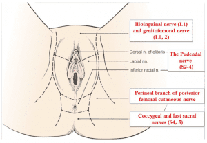
In my practice, I define it as a pain located in the area of innervation of the pudendal nerve. Pudendal nerve entrapment is an impingement of the pudendal nerve caused by scar tissue, surgical materials, or mesh. Pudendal nerve entrapment is, therefore, one of the causes of pudendal nerve neuralgia, however other causes such as inflammation, spasm of the surrounding muscles, or other nerve diseases may also be a reason for pain.
Pudendal nerve entrapment is almost always caused by some traumatic event to the pelvis. This may be pelvic surgery (with or without mesh), difficult childbirth, athletic injury, falls, and other accidents. A repetitive injury such as bicycle seat pressure on the pelvic floor may also lead to pudendal nerve entrapment (cyclist syndrome).
Diagnosis of pudendal nerve entrapment is not easy and relies heavily on taking a detailed history. Pain is located in the vagina, vulva, clitoris, perineum, and rectum, and it may involve one or all of those areas. Pain is more severe with sitting than with standing or laying down and sitting on the toilet is generally better than sitting on the chair. Most of the patients with real nerve injury have pain on one side only, or one side is significantly more painful than the other. Pain is generally more severe with urination, bowel movements, and intercourse. Some patients may also have difficulty emptying their bladder (hesitancy) and bowel (constipation). One of the most debilitating symptoms of pudendal nerve entrapment is a sensation of continuous sexual arousal (persistent genital arousal disorder – PGAD). Patients often reduce this sensation by masturbation which only provides temporary relief.

A pioneer in the treatment of pudendal nerve entrapment and my mentor, Professor Roger Robert, has developed Nantes criteria which greatly assist in diagnosing this condition. Studies have shown that patients who more closely meet the criteria have better outcomes from the surgical decompression of the nerve.
Pain in pudendal nerve entrapment is of neuropathic nature, which means that patients living with pudendal neuralgia feel burning tingling and numbing sensation (paresthesia). Some patients have the sensation of a foreign body located in the rectum or vagina (allotriesthesia) and may describe it as a “red hot poker” in the rectum. Some patients do not experience any pain but have complete or partial numbness in the area of innervation of the pudendal nerve.
Additional tests such as magnetic resonance neurography (MRN), pudendal nerve motor terminal latency (PNMTL), another electrophysiologic testing, or sensory threshold testing are generally not accurate enough to diagnose pudendal nerve entrapment. A CT-guided pudendal nerve block is a part of Nantes criteria, and an important step in the diagnosis and treatment of pudendal nerve entrapment. Lack of relief of pain immediately after a CT-guided pudendal nerve block means that pain originates in another structure or is transmitted by a different nerve other than pudendal.
Conservative treatments of pudendal neuralgia consist of:
- Avoidance of additional injury – patients need to immediately stop the activities that lead to injury of the nerve in the first place. For example, if nerve pudendal neuralgia was caused by riding a bicycle, the patient has to immediately stop cycling. Of course, this cannot be done in cases where the patient developed pudendal neuralgia as a result of surgery or childbirth
- Protecting the nerve by using sitting cushions, zero gravity chairs, or kneeling chairs
- Medications including oral medications and vaginal/rectal suppositories
- Appropriate pelvic floor physical therapy (to minimize pelvic floor muscle spasm)
- Botulinum toxin A injections to pelvic floor muscles (to minimize pelvic floor muscle spasm)
- Pudendal nerve blocks using CT, ultrasound, or in some cases unguided transvaginal blocks
- Pudendal nerve injections with amniotic fluid and liquified amniotic membrane
- Ablation procedures – pulse radiofrequency ablation (pRFA) and cryoablation
- Nerve stimulators and spinal cord stimulators
- Surgical decompression of the nerve. Pudendal nerve decompression can be done using several different approaches: transgluteal, transischorectal, transperineal, and laparoscopic/robotic.
The transgluteal approach is an original technique described by Professor Roger Robert in Nantes, France, and very significantly modified by me. This approach offers by far the best access to the pudendal nerve, therefore allowing for the most complete decompression. One of the earlier drawbacks of the technique was cutting of the sacrotuberous ligament which in some cases could lead to pelvic instability. The risk of that instability was eliminated when I began repairing the sactotuberous ligament. Cutting off that ligament allows access to the nerve and freeing it from the scar tissue or surgical materials, but after nerve decompression is accomplished sacrotuberous ligament should be repaired.
Other modifications that I have introduced to the pudendal neurolysis surgery were:
- Use of surgical microscope for better visualization of the nerve and surrounding structures
- Use of Nerve Integrity Monitoring System (NIMS monitor) to aid with identification of pudendal nerve. It is especially helpful in cases when the nerve is significantly scarred, and in cases of repeat surgery.
- Use of a pain pump that delivers a local anesthetic to the nerve for about seven days after surgery. This decreases pain levels and is thought to reverse central sensitization (memory of pain in the brain). This step may lead to faster recovery and resolution of pain after surgery.
- Nerve wrapping with an adhesion prevention barrier decreases the risk of scarring or re-scarring of the nerve after surgery. Several years ago, I switched from regular nerve wraps to wrapping the nerve with an amniotic membrane product. In addition to anti-adhesion (anti-scarring) properties, the amniotic membrane contains nerve growth factors that promote nerve healing. It also may have the ability to attract your own body’s stem cells close to the nerve, which further helps with nerve regeneration.
- Use of suction dressing after the closing of the skin to minimize the risk of wound infection.
Laparoscopic/robotic procedure is less invasive, but it does not offer as good of access to Alcock’s canal as transgluteal procedure does. It may be effective in cases where the nerve compression is limited to the small area, but the location of compression may be difficult to determine prior to surgery.
In my practice, I perform both transgluteal and robotic nerve decompression procedures, but I believe that since transgluteal technique offers better access and allows for decompression of the larger part of the pudendal nerve, it should be a preferred approach. Even though the recovery time is longer compared to the laparoscopic procedures, the benefits of more complete nerve decompression are very important when considering the choice of surgery.
Overall, the results of surgery show that the majority of patients have significantly decreased pain, and benefit from pudendal nerve decompression procedures.
If you or someone you know experiences pain with sitting in the clitoris, vulva, penis, scrotum, perineum, or rectum, call 480 599-9682 or email our clinic at [email protected] to learn more about available treatments.
What to expect before and after transgluteal nerve decompression surgery?
1. Prior to surgery Dr. Hibner and his team will use some or all available non-invasive methods to help you with your pain. They may include physical therapy, suppositories, oral medications, nerve ablations, injections of amniotic fluid/membrane products.
2. Decision for the surgery is made together by the patient and Dr. Hibner. It is based on all the information from the patient’s history, exam, radiology results, any additional testing. Also, the fact that all offered conservative treatments have failed is taken into account when deciding on surgery.
3. Transgluteal pudendal nerve decompression is Dr. Hibner’s preferred way to free up the nerve from scar tissue but in certain cases, a robotic approach or highly selective approach to pudendal nerve branches may also be chosen.
4. Prior to the surgery please follow all the pre-operative instructions which will be given to you during the visit
5. For the transgluteal decompression surgery you will be positioned on your abdomen (prone) and the incision will go on the buttock on the operated side. It will measure anywhere from 2 to 4 inches.
6. When you wake up from surgery in the recovery area you will have a catheter in your bladder, a pain pump dripping local anesthetic on your nerve, and negative pressure dressing on the skin over the incision. You will receive a bag to wear over your shoulder for the pain pump and device providing suction to the dressing). It is very important to be gentle with the pump to avoid dislodging the catheter. If the pain pump catheter is dislodged, it cannot be replaced.
7. You should feel numbness in the area of the pudendal nerve (where the pain was before surgery) but the incision over the buttock will be quite tender. Pain pump medication does not reach the muscles or skin of the buttock, and it is only meant to provide pain relief in the nerve.
8. Most patients spend one night in the hospital. Rarely, a two-night stay is required
9. You should be active and try to walk with support the next day after surgery. This prevents a loss of muscle and could also decrease the risk of nerve scarring. Please be very careful when moving and walking not to pull the pain pump.
10. You will be discharged home with pain medications and instructions on how to use the pain pump and negative pressure dressing. You may need to return to the clinic for the dressing and pain pump removal, or we will instruct you how to do it yourself. When the pain pump comes out your pain may increase until the nerve begins healing.
11. You can shower 2 days after surgery. Try to avoid making the area of the incision wet. You can wrap that area with a large garbage bag for the time of surgery. Water is not going to contaminate the incision but may make the adhesive covering the pain pump catheter wet leading to the earlier removal of the pump.
11. Most patients can travel within 5-7 days after surgery but a longer stay is encouraged before traveling back home.
12. You can resume the activity after surgery but avoid doing things that significantly increase your pain. Sitting should be avoided. If surgery was done on one side only try to sit on the opposite buttock.
13. Do not flex your hip(s) over 90 degrees. This may lead to pulling apart the sacrotuberous ligament which was repaired during surgery. This ligament takes approximately 6 months to heal. You should therefore avoid squatting, taking two steps at a time when walking upstairs, etc.
14. You should resume pelvic floor physical therapy approximately 6 weeks from surgery. You will be given instructions for you and your physical therapist regarding recommended therapy.
15. It may take 3-4 months to start feeling the improvement in pain. It is normal to have better and worse days. When you have a better day please be careful not to overexert yourself. Follow the instructions of your pelvic floor physical therapist on physical activity.
16. Please continue to take all your medications until you are instructed to stop. You can continue vaginal/rectal suppositories, but you should be decreasing the number of narcotic pain medications you take.
17. Generally we do not schedule patients for in-person postoperative visits. A majority of care can be done over the phone or via telemedicine.
18. Maximum healing may take 18 to 24 months from surgery. After 2 years from surgery majority of patients have less pain or no pain. If you continue to be in pain, we will look for other solutions to help you with your condition.
Outcomes – Does Pudendal Neuralgia go away?
Outcomes of this procedure depend on the causes of nerve compression, the degree to which the nerve was compressed, and how much time elapsed between the injury and surgery. Unfortunately, the degree of nerve damage is difficult to assess before surgery. From my extensive experience of doing hundreds of pudendal decompression surgeries, approximately two-thirds (66%) of patients benefit from this procedure. This number includes all the patients, even those with severe nerve injury. That means that patients with less severe nerve injury may benefit from the procedure even more.
Pudendal Neuralgia History…
Pudendal neuralgia has been recognized by medicine in the book published in Philadelphia in 1871 – “The Change of Life in Health and Disease”. The knowledge of pudendal neuralgia was almost lost until the late 1980s. French neurologist Dr. Gerard Amarenco reported on a series of patients with “syndrome du cyclist”, the cyclist syndrome which occurs when a pudendal nerve is compressed between narrow bicycle seat and medial surface of ischial tuberosity (sitz bone). The first procedure to surgically decompress the pudendal nerve through transperianal technique (incision around the anus) was described in 1992 by Egyptian surgeon Dr. Ahmed Shafik.
Soon after my mentor Professor Roger Robert from Nantes, France described transgluteal pudendal neurolysis – decompression of the pudendal nerve with an approach through the buttock. Professor Robert is not only an outstanding neurosurgeon, but also an anatomist, and this unique combination allowed him to develop the whole new procedure for pudendal nerve decompression.
I graduated from my fellowship in gynecologic surgery at Mayo Clinic in 2003 and opened pelvic pain practice in Phoenix in 2004. I started seeing patients with pelvic pain whose condition could not be explained by any disease known to me. So, in early 2005 I googled the symptoms: perineal/vaginal bringing pain with sitting. Several medical articles showed up, but most of them had one common name as one of the authors: Roger Robert. I then send the letter to Nantes France to Professor Robert if I could come to visit him and learn from him.
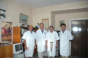
In the summer of 2005, I traveled to Nantes and worked with Professor Robert for almost 3 weeks assisting him on numerous pudendal neuralgia surgeries and seeing many patients in the office with him. I also worked with wonderful Dr. Jean Jacques Labat, a neurologist who assisted Professor Robert with diagnosing and treating patients with pudendal neuralgia before surgery, and with amazing radiologist Dr. Thibault Riant who taught me how to perform CT-guided pudendal nerve blocks.
When I returned to Phoenix, I started seeing more and more patients with pudendal neuralgia and pudendal nerve compression, and I performed my first transgluteal pudendal nerve decompression in the fall of 2005. It was in the patient who developed pudendal neuralgia after removal of Bartholin’s gland. She did well after surgery and soon many more patients have followed.
From the very first surgery, I started modifying the original procedure developed by my great mentor, Professor Robert. The first modification was repairing the transected sacrotuberous ligament. There was a concern that leaving this ligament not repaired may cause instability in the sacroiliac joint. So, from the very first patient, I would repair sacrotuberous ligament the same way that an orthopedic surgeon repairs a ligament in the knee. Next, I incorporated the use of a neurosurgical microscope into the procedure. This allowed for significantly improved precision.
The next modification was the use of an On-Q pain pump placed next to the nerve towards the end of surgery to provide postoperative analgesia and decrease central sensitization (memory of pain in the brain). The third modification was the incorporation of NIMS (nerve integrity monitoring system) to allow the identification of the nerve in the setting of significant scarring. The next modification was the use of nerve wrap to prevent the reoccurrence of adhesions.
Initially, I was using a collagen nerve conduit but a few years ago I switched to the amniotic membrane which in addition to preventing adhesions also contains factors/chemicals promoting nerve healing. The last major modification was the method in which I cut sacrotuberous ligament. Cutting it in a Z fashion allows me for better access to the nerve and facilitates the repair at the end of surgery.
Up to today, I have done several hundred of those procedures, most likely more than any other provider with exception of my amazing mentor, Professor Roger Robert.

Special thanks
Hard work and knowledge of many, many people went to the development of transgluteal pudendal decompression surgery the way I perform this procedure today. I would like to take this opportunity and thank: Professor Roger Robert, Professor Oskar Aszman, Dr. Jamie Balducci, Dr. Jacek Bendek, Dr. Mario Castellanos, Dr. May Nour, Cindy Love, and many others.
Big thank you from me and on behalf of my patients whom I was able to help with pain management.
